 Round window - Wikipedia
Oval WindowRelated terms: Francis A. Spelman, in , 2013 The Periphery The hearing periphery consists of the external ear, the middle ear and the inner ear. The external ear, the pine, collects changes in the pressure (condensations and rare productions) produced by the hearing signals. These signals are generated by sound sources in the listener's environment. Acoustic signals are guided to the middle ear along the ear canal, an entry into the head of the subject that is lined with soft tissue. The auditory canal is open at its peripheral end, and tied by the eardrum, the tyrannical membrane, at its inner end. The length of the ear canal is about 3 cm in the human (Geisler, 1998). The middle ear is tied distally by the tymponic membrane and next by the claw, where the footplate of the staps is contacted with the oval window of the claw.1 In the middle ear there are three small bones that make up the osicular chain: the malleus (hammer), incus (anvil), and stapes (stirrup). Bones have flexible connections. They provide a mechanical advantage so that the eardrum can be driven by the air and, in turn, can drive the dense liquids found in the cochlea. The movement of the standing plaque of the staples is about 75% of that of the tympanic membrane in the human (Geisler, 1998). Other mechanical advantages are the relative areas of the tymponic membrane and the footplate of the staves: the tymponic membrane has about 20 times the area of the staves (Geisler, 1998). Small pressure changes in the air are transformed into greater pressures in the cochlea fluids. On the contrary, the large displacements of the eardrum produce small displacements of the standing plate of the stapas. Cochlea is a snail-shaped organ that is bilaterally found in the temporary bones of the head. The cochlea is oriented in such a way that its broad base faces in a medium and later direction, while the axis of its lateral and previous spiral points (Figure II.5.11.1). The cochlea contains three spiral or scalae chambers: the tympani scala, scala vestibuli and scala media. The organ of Corti is within the means of scala in the basilar membrane. The capillary cells are found in the basilar membrane (Dallos et al., 1996; Geisler, 1998). The basil membrane is an elegant structure that acts as a Fourier mechanical analyzer; specific regions of the membrane vibrate to the maximum in response to the frequency of the sound waves imposed on the staves. Membrane displacements produce maximums for high frequencies in the basal end and for low frequencies in the apical end of the cochlea. The capillary cells that reside in the membrane have cilias that are bent when the membrane vibrates. The hair cells are synapsed with the peripheral processes of the auditory nerve, the auditory nerve in Figure II.5.11.1. There are 25-300,000 aferent neurons that are synapsed with the hair cells (Geisler, 1998). The organization of the auditory nerve is due to the frequencies of the acoustic signals it receives. In fact, the entire auditory system is organized tonotopically, i.e., by frequency (Popper and Fay, 1991; Geisler, 1998). Tonotopic Account System Organization The entire auditory system analyzes the frequency of the signals it receives. The peripheral system begins the process (Geisler, 1998), and that the anatomical organization proceeds at least to the level of the lower colculus, in the brain medium (Snyder et al., 2004). As illustrated dramatically by Biererer, the organization can be detected in the auditory cortex (Bierer and Middlebrooks, 2004). This organization allows researchers to test the effectiveness of animal simulation designs (Bierer and Middlebrooks, 2002, 2004; Snyder et al., 2004). His work has allowed others to design and test medium brain implants in human subjects suffering from hearing nerve tumors (Lim et al., 2008). While the tonotopic organization of the auditory system is a global part of its design, many subtleties, for example, the temporary analysis of the fine structure, accompany the organization frequently. These subtleties seem to underestimate the ability of the system to extract sound signals and decode music (Rubinstein et al., 1999; Litvak and Narayan, 2007; Wilson and Dorman, 2008). Critical to the design of the cochlear prosthesis is the location of the peripheral processes of the auditory neurons, the cells of the audity or hearing nervio.2 Neurons are bipolar cells, and their peripheral processes are found inside and under the boní spiral lamina (Popper and Fay, 1991; Spelman and Voie, 1996; Geisler, 1998), a bold or bold structure that extends from the modium (medial) wall of the tympani scala in the cochlea. The cell bodies of the VIII nerve are located in the Rosenthal canal, a spinal structure in the Modium bone. The anatomy of the scala tympani has led to the design of the cochlear implant. Prosthesis is designed to stimulate auditory neurons electrically. Placing electrode sites near neurons without violating the modiolus bone wall requires the electrode arrays of the implants to be located in the tympani scala (vide infra). Jeffrey David Lewine, in , 1995Auditory and Vestibular Systems40 The hearing system is a complex system designed for precise characterization of sound intensity and field. A series of mechanical elements, the osicles middle ear, transmit vibratory energy related to sound from the eardrum to the oval window, where vibrations are established in the fluid media of the cochlea (Fig. 2-72). The organ of Corti, located on the upper surface of the cochlea basilar membrane, contains specialized cells of hair that act as the neuronal transducers in this system. Basil membrane oscillations produce an activating force in the cilia of the hair cells. Fig. 2-72. A, the peripheral and vestibule hearing structures are inertiated by the eighth cranial nerve. B, The middle and inner ear with schematic vision straight from the cochlea. C, Extended view of the section through the cochlear duct that illustrates the Corti organ. The mechanical properties of the basil membrane are such that the high frequency sounds cause oscillations at the base of the membrane, near the oval window, while the low frequency sounds cause oscillations in the apex. This imparts tonotopic organization to capillary cells throughout the Corti organ. Apex cells encode low frequency sounds and base cells, high frequency sounds. When the cells of the hair are activated, they release neurotransmitters that activate the peripheral axes of the spiral nodes cells that invade them. The central projections of the spiral ganglia cells form the cochlear branch of the cranial nerve VIII, the coibulococlear nerve. The Fig. 2-73 provides the details of the central route of projection to the primary auditory cortex of the transverse temporal spin (Heschl's) , Brodmann area 41. The tonotopic organization of primary hearing projection fibers is maintained along the projection path. Neurons along Heschl's spin best respond to high-frequency tones while neurons close to the lateral margin of the spin respond better to low-frequency tones. The primary hearing cortex projects several regions of association, including Brodmann area 22 and Wernicke area. These are found along the dorsal and lateral aspects of the upper and middle temporal spin. Transcallosal projections are also prominent. Fig. 2-73. A, Central hearing system projections. B, Central protections of the vestibular system. It should be noted that several complex hearing computations are performed in early subcortical cores in the hearing processing track. The role of the superior olive complex in computations related to sound location should be highlighted. The vestibular apparatus, located in the inner ear along with the cochlea, includes semicircular channels, the utricle and the sacrcle (Fig. 2-72). The specialized hair cells, similar to those found in the cochlea, are the receptor cells of the vestibular system. These cells are inervaded by the distal process of neurons located in the Scarpa nodes. The central projection axes of these neurons form the vestibular division of the cranial nerve VIII. Figure 2-73 illustrates the central pathways of the vestibular system. The vestibular cores of the brain trunk contain the critical circuit for several vestibule reflections, including the very important vestibular-oculomotor reflections. The cortical representation for the vestibular function is poorly defined, but on the basis of connectivity, the relevant cortical region is believed to be in the parietal lobe only flow to the primary somatosensor cortex. Joseph Feher, in , 2012Pitch's perception is completed by a combination of phase tuning and blockAs described above, the main fans for sound detection emerge from the inner hair cells that respond to the deformation of the stereos in response to the movement of the tectorial and basilar membranes. Pressure waves transmitted to the oval window through the action of the osicles produce a wave of basilar membrane deformation that has a maximum displacement that depends on the frequency, but also depends on the magnitude of the pressure waves. The physical response of these membranes, along with the electrical properties of the inner hair cells, produces a "learning curve" for primary afferent fibers. These tuning curves are the threshold intensity for the generation of impulses as a frequency function. Typical tuning curves for primary auditory nerve aferents are shown in Figure 4.7.8. Figure 4.7.8. Adjustment curves for primary aferents in different locations in the cochlea. The afferents of the inner hair cells near the apex show maximum sensitivity (the lower threshold) to the sounds with low frequencies (black curve). The primary afferents of the inner hair cells near the base show maximum sensitivity to the higher frequencies (sight at the far right). Cells between the intermediate behavior of the show (medium two curves). Sensitivity falls sharply into higher frequencies, but the lower frequencies can still excite the afferents of their maximum sensitivity. The tuning curves show a fairly sharp maximum sensitivity (the lower threshold) to a characteristic frequency. Sensitivity falls sharply into higher frequencies, but usually shows a "tail" in lower frequencies. This is because itinerant wave for high frequencies travels up to the moment to the cochlea, while all low-frequency sounds should pass through the location of the cells of the inner hair to reach their place of maximum displacement of the basilar membrane. The higher sounds produce larger deformations of the basilar membrane, so a stronger sound can be confused with one of greater tone. There is psychophysical evidence for a change in the field perceived with sound intensity. The auditory system deals with this problem using a sound frequency phase block encoding as well as the adjustment curves of the primary afferents. Because a pure tone transmitted to the perilymph induces a sinusoidal displacement of the basilary membrane, primary aferent neurons tend to shoot in phase with the sound wave. Therefore, neurons shoot the same part of the sound wave, usually their maximum pressure. Therefore, your shooting rate encodes the original sound frequency. However, each primary afferent has a limited shooting rate of less than 200 Hz; they cannot keep up with the tones that have a higher frequency than this. Because each inner hair cell is inervated by 10 or more primary afferents, the set does track the stimulus waveform. The convergence of these primary afferent neurons in objectives within the cochlear nucleus suggests that the summation within the cochlear nucleus allows a tonometry frequency coding and a place encoding (the tonotopic map). This requirement probably explains the curious multiple inervation of the inner hair cells. These combined codes allow simultaneous discrimination of intensity and sound tone. Kuang-Hua Chang, in , 20157.6.1 Case study In this case, we present a practical and systematic method of building a precise finite element model for a human middle ear (Sun et al., 2002). The middle ear is the portion of the inner ear to the eardrum and external to the oval window of the cochlea, as shown in Figure 7.38. It contains three oses (malleus, incus and stapes), which transform the vibrations of the eardrum into waves into the fluid and membranes of the inner ear. The hollow space of the middle ear is the timponic cavity, or the tympani cavum. The eustachian tube joins the timpian cavity with the nasal cavity (nasopharynx), allowing the pressure to equal between the middle ear and the throat. Figure 7.38. Half-human ear anatomy. The components of the middle ear have a small and complex geometry, and play an essential role in sound transmission. The sounds collected and transported in the external auditory channel are first transformed into mechanical vibrations of the eardrum and ossicular chain, and then transformed into itinerant waves in the coclea filled with fluids (inner order). Devices for hearing restoration in cases of medium ear (sensory loss) impairment are often mechanical in nature and can therefore be studied using mechanical computational models. From the perspective of mechanics, the middle ear is essentially a coincidence of impedance between the external ear and the inner ear. Its transfer function, related to the dynamic behavior of the osicular chain, is complex due to its anatomy. The aim of this case study was to create a precise three-dimensional geometric model of the human middle ear that allows to observe its 3D structure for morphological studies and to develop models of finite elements for mechanical analysis. The modeling process began with the extraction of a normal cool human temporary bone that was fixed, disqualified, and encrusted in jealousyidine. The resulting block was removed from the chloroform, trimmed, mounted on a microtome plate, and stored in 70% of the alcohol. Before the block of the jealousy was dried, four parallel fiducial holes (perpendicular to the cutting plane) were made in it with a drill press. Permanent ink is injected into fiducial holes to stain the marks. The sections were cut into a slide microtome at a thickness of 20 μm, as shown in chapter 2, Figure 2.36(a). Then, in sequence, each tenth section was stained and mounted on glass slides. Finally, each histology slide was scanned on a computer using a flat-bed scanner and saved as an image file (chapter 2, Figure 2.36(b)). The images were aligned with a template built by a typical section image using fiducial marks. The images of the alignments were cut as standard-size images, taken to a sketch plane in the CAD software (in this case SolidWorks), and fitted into a reference rectangle of the same size as the image so that the actual size could be accurately represented. The images were then digitized by marking points along the outlines of the medium ear structures, as identified by an otological surgeon. These structures included eardrum, osicles, attachments and muscle tendons (chapter 2, Figure 2.36 (c)). The B-spline curves were built to better adapt to these points using the curve adjustment technique (chapter 2, Figure 2.36(d)) and were paved to form the closed boundary surfaces for the individual components of the middle ear using the surface skin technique (Tang and Chang, 2001). The solid models of the components of the middle ear were built using these surfaces and then assembled to create the whole model of the middle ear, as shown in chapter 2, Figure 2.36(e). All the surfaces of the geometric model were translated into HyperMesh for the modeling of finite elements. Based on these surfaces, the FE mesh of the components of the middle ear and attachments/tendons was created with HyperMesh machining capabilities. Chapter 2, Figure 2.36(f) shows the previous-medial view of the mid-eared FE model with attachments/tenes and cochlear fluid limitations. A total of 1,746 triangular elements of three knots and four knuckles of quadrilateral shell were created to mesh the eardrum. Around the periphery of the eardrum, the timponic anulus was modeled using 113 triangular elements of three knots and quadrilaterals of four knots. A total of 812 hexahedral of eight nodes, pentahedral of six nodes, and solid tetraedral elements of four nodes were created for mesh three oscillales, two joints (incudomalleolar and incudostapedial pairs), and six previous ligaments/tendons (superior mallearment C1, mallearment C2, posterior). Lying on the floorboard plane, 25 spring elements were used to model the anular stapedius ligament. A total of 49 perpendicular elements to the floor plate plane were incorporated to model the cochlear fluid (coclear impedance). The total number of nodes was 1,497 and the number of degrees of freedom was 4,491 in the model shown in figure 2.36 (f). The size of the model was suitable for a FE analysis with a reasonable calculation time. The lynxes of the FE model included suspensory ligaments, intra-aural muscle tendons, timponic anulus, stapedius anular ligaments and cochlear fluid. As shown in Figure 2.36(f), the malleum, incus and staples are attached to the eardrum by the malleum and in the window oval by the pieplate of the staves. Suspensory ligaments and intraaural muscle tendons also support the oses. Four main suspensory ligaments (superior malleus C1, lateral malleus C2, posterior incus C3, and anterior malleo C4) and two intraaural muscle tendons (postal C5 and tensor tympani C7) were considered as elastic restrictions. The timponic annulus was modeled as an elastic ring that connects the eardrum periphery and the bone wall of the ear canal. The cochlear fluid was assumed as a viscoelastic limitation, C6. Frequency response analysis was performed using ANSYS. An excellent resolution of the displacements of the standing plates in six human temporary bones was observed using Doppler laser interferometry on the ranges of acoustic frequencies, including 80, 90 and 100 SPL acoustic inrush in the timpan membrane. The results of the finite element model show that the 3D model of the human middle ear is suitable for the investigation of the medium ear mechanics and the transfer functions. A.A. Kuznetsov, O.A. Kuznetsov, in , 20033 Protecting the median ear sound behavior system after radical surgeryIn patients with median ear sound drive system completely destroyed, but an intact inner ear, hearing rehabilitation requires both the adjustment of the impedance of the prosthesis with the impedance of the inner ear and the round window shielding. Magnetic soft ferrommetic particles (steel X13) were implanted under the scar tissue of the oval window area in postoperative patients with chronic supurative otitis media, waterproof hearing tube and total absence of the sound conduction system (Kuznetsov et al. 1982, Palchun et al. 1982). A removable elastic confusor, which contains small permanent magnets, was attached to the claw window by magnetic forces (Fig. 1). This confusor focuses the sound pressure in the oval window while protecting the round window. Since the magnetic forces guide the confusor to the proper position during insertion into the auditory channel, this procedure can be done by the patient. Confusor protector provided up to 35 dB decrease in air conduction thresholds in 18 patients, which is approximately twice better, than traditional techniques such as round window armor by cotton tampons with oil or silicone foam "open" (Wullsten et al. 1972, Williams and Rouf 1978). Richard Brice, in , 2003 Ear PhysiologyStudies of the physiology of the ear reveal that the Fourier analysis process, referred to above, is more than a mere mathematical conception. Anatomical and psychophysiological studies have revealed that the ear performs something very close to a Fourier mechanical analysis on the sounds it collects, and passes a frequency domain representation of those sounds to higher neuronal centers. Figure 2.7 contains an illustration of the human ear. Figure 2.7. The physiology of the earAfter interacting first with the auricle or pine, the sound waves run through the ear canal to the eardrum. The position of the eardrum marks the limit between the external ear and the middle ear. The middle ear is an air-filled cavity that houses three small bones; the hammer, the anvil and the fuss. These three bones communicate the vibrations of the eardrum to the oval window on the surface of the inner ear. Because of the way these bones are pivoted, and because the base of the hammer is wider than the base of the fuss, there is a considerable mechanical advantage from the eardrum to the inner ear. A tube runs from the base of the middle ear to the throat; this is known as the Eustachiano tube. Its action is to ensure that there is equal pressure on both sides of the eardrum, and it is open to swallowing. The inner ear is formed in two sections; the claw (the spiral structure that looks like a snail shell) and the three semicircular channels. These latest structures are involved in the sense of balance and movement. The stirrup is firmly linked to the membrane that covers the opening of the oval window of the claw. The cochlea is filled with liquid and is divided throughout its length by the Reissner membrane and the basilar membrane, on which the Corti organ rests. When the stirrup moves, it acts like a piston in the oval window and this puts the fluid inside the cochle in motion. This movement, trapped inside the closed ball, creates a permanent wave pattern – and therefore a distortion – in the basilar membrane. It is important that the mechanical properties of the basil membrane change considerably along its length. As a result, the position of the peak in the vibration pattern varies depending on the frequency of stimulation. The bearing and its components work as well as a frequency-to-position translation device. Where the basil membrane is diverted further, there are the cells of the organ of Corti; these interfaces the aferent neurons that carry signals to the upper levels of the auditory system. Therefore, the signals coming out of the ear are in the form of a frequency domain representation. The intensity of each frequency range (the exact nature and the extent of these ranges are considered later) is encoded by a pulse rate modulation scheme. Joseph Feher, in , 2012 The chapter begins with a description of the subjective experience of hearing: tone, bell and noise. The anger is measured in decibels in relation to a standard. The structure of the ear is checked: external ear, middle ear and inner ear. The timponic membrane impedance system, oses, oval window and cochlear fluid is discussed. The structure of the cochlea and the organ of Corti is described. Multiple inervation of the inner hair cell row is contrasted with multiple efferent inervation of the triple outer hair cell row. The tonotopic cartography of the cochlea and its preservation are discussed along the path to the hearing cortex within the lateral fissure or Sylvian sulcus. The sound transduction mechanism is also discussed, with the peculiar high composition [K+] and the positive potential of endolymph and the pointed links that bind kinocilium to the other stereos. The path that leads from the spiral node cells to the primary hearing cortex is drawn. Recommended Publications: We use cookies to help provide and improve our personalized service and content and ads. By continuing to accept . Copyright © 2021 Elsevier B.V. or its licensors or collaborators. Direct Science ® is a registered trademark of Elsevier B.V.ScienceDirect ® is a registered trademark of Elsevier B.V.
Oval Window The human ear consists of three regions called external ear, middle ear and inner ear. The oval window, also known as the fenestra ovalis, is a conjunctive tissue membrane located at the end of the middle ear and the beginning of the inner ear. The fenestra ovalis connects the small bones of the middle ear to the vestibuli scala, which is the upper part of the cochlea. (The claw is the central organ of the inner ear.) The bone of the middle ear that really connects to the fenestra ovalis is called the fuss, or stapes. The middle ear works to transmit the movement of the eardrum (or the timpan membrane) to the inner ear. This increases the pressure on the connective tissue of the oval window. This pressure is ultimately transmitted through the stakes, which press against the fenestra ovalis, to the cochlea. From there, it travels through the auditory nerve to the brain, which processes the sound. Last medical review on January 21, 2018Read this following

oval window ear model - Google Search | Ear infection remedy, Ear infection, Ear infection home remedies
Ear Model.mov - YouTube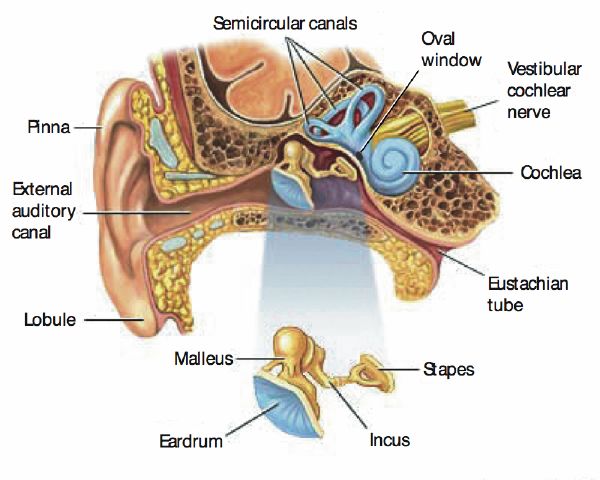
Tristan Jehan PhD Thesis - Chapter 3
Eardrum - Wikipedia
Inner-Ear Barotrauma (IEBT) | Ears & Diving - DAN Health Issues & Diving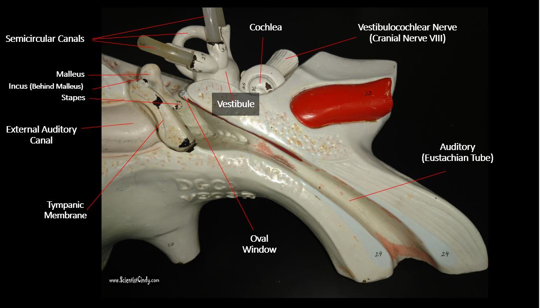
The Ear - Sensory Organs - SCIENTIST CINDY
bone structure model labeled - Google Search | anatomy | Pinterest ... | Ear anatomy, Anatomy models labeled, Anatomy models
2. Anatomy of Ear - msn
Round window | anatomy | Britannica
Structure of the Ear: How the Ear Works | Beltone South
Ears and Eyes Anatomy Flashcards | Quizlet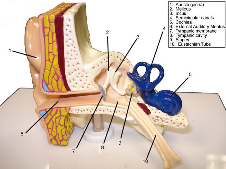
Lab Guide: Ear, Eye, Brain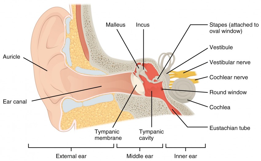
Audition and Somatosensation | Anatomy and Physiology I
Solved: Label The Following THE EAR Auditory Eustachian Tu... | Chegg.com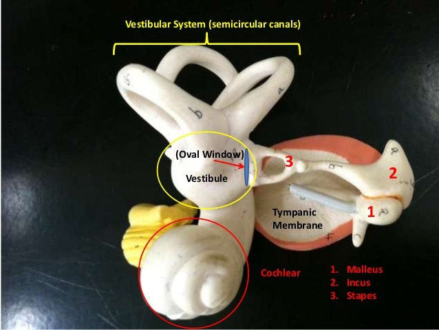
Ear models/GettyImages-506837613-579d8e123df78c3276da6870.jpg)
Symptoms and Causes of Perilymph Fistula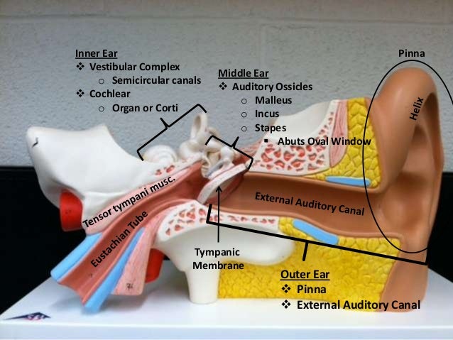
Ear models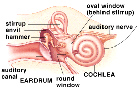
oval window
Middle ear - Wikipedia
Ear Diagram Oval Window - Human Body Anatomy
Incus (Anvil) - an overview | ScienceDirect Topics
Ear model - YouTube
Round window | anatomy | Britannica
Osseous labyrinth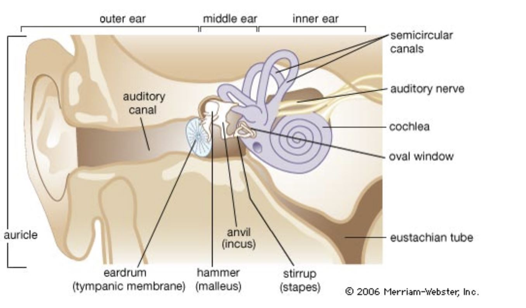
Solved: A Person's Ear Is Measured To Have A Middle Ear Pr... | Chegg.com
CH 15 Inner Ear - Hearing
The Anatomy of The Human Ear – Empire Ears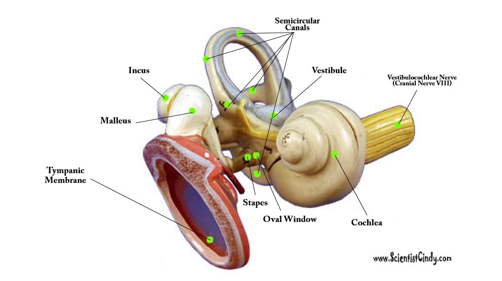
The Ear - Sensory Organs - SCIENTIST CINDY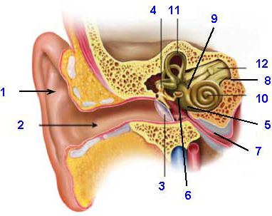
The Normal Ear - Understanding Parts of the Ear and How We Hear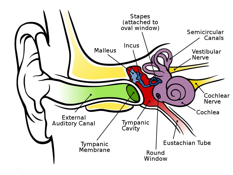
How does our hearing work and how is the ear structured?
Anatomical barriers of the ear (RWM: Round Window Membrane). | Download Scientific Diagram
Oval Window Ear Model Labeled (Page 1) - Line.17QQ.com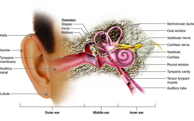
Hearing and Equilibrium | Anatomy and Physiology
ear model | Ear anatomy, Human anatomy and physiology, Anatomy
The Sense of Hearing - The Nervous System: B. The Special Senses - Guyton and Hall Textbook of Medical Physiology, 12th Ed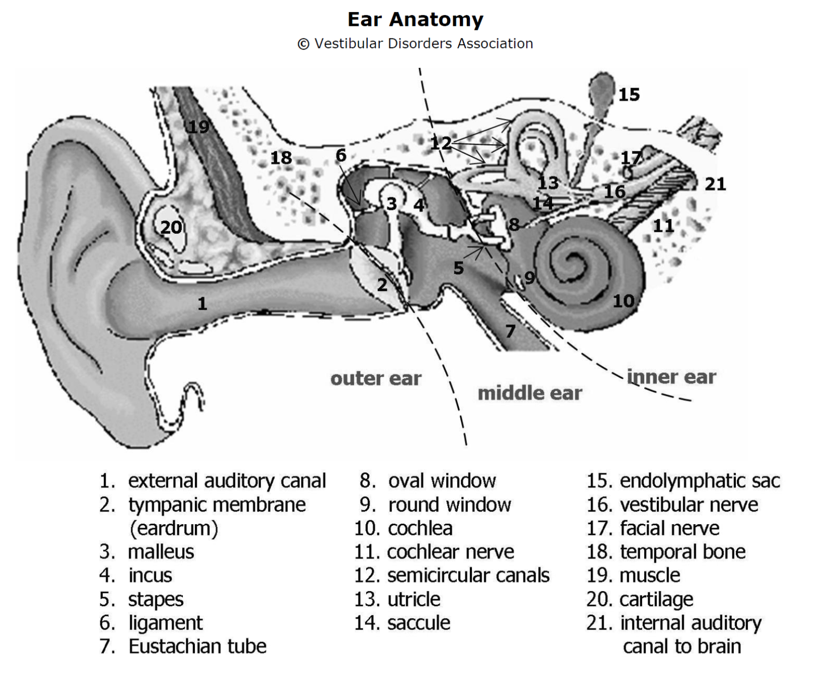
Ear Anatomy
Biomechanics of Human Hearing | Audio Science Review (ASR) Forum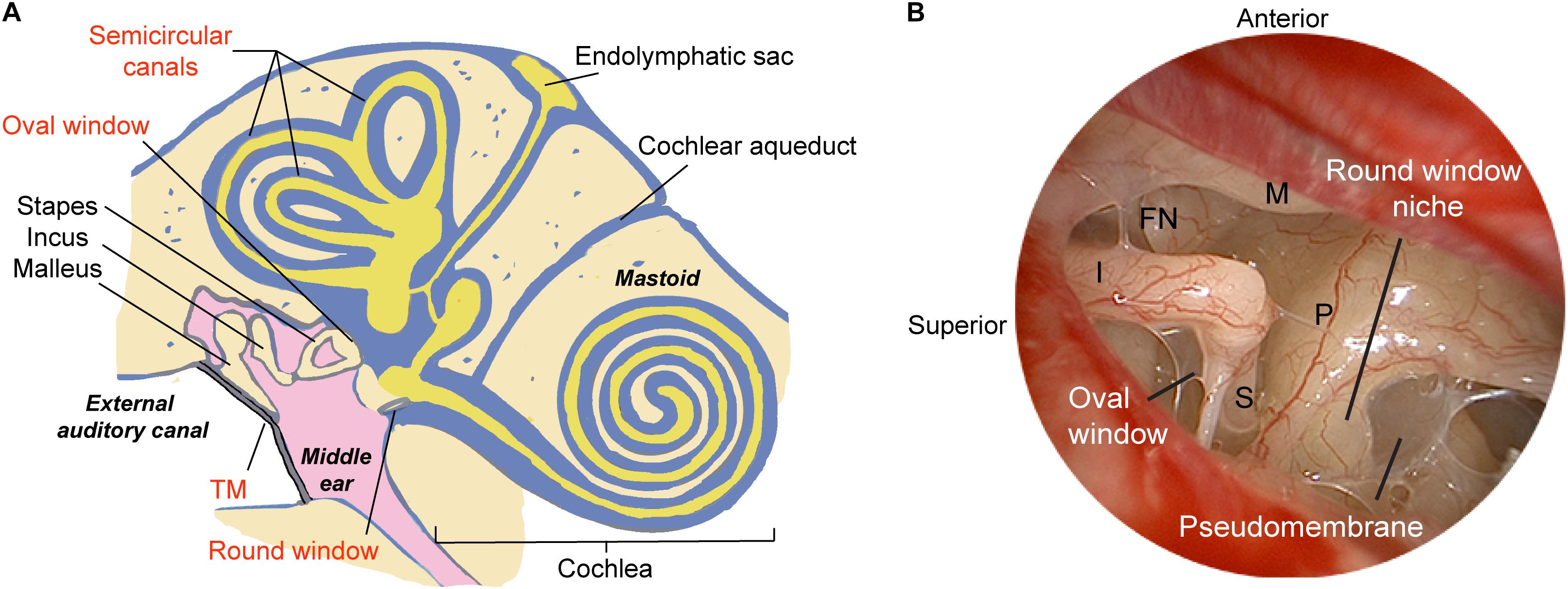
Frontiers | Gene Therapy for Human Sensorineural Hearing Loss | Cellular Neuroscience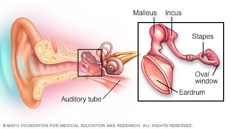
Slide show: How you hear - Mayo Clinic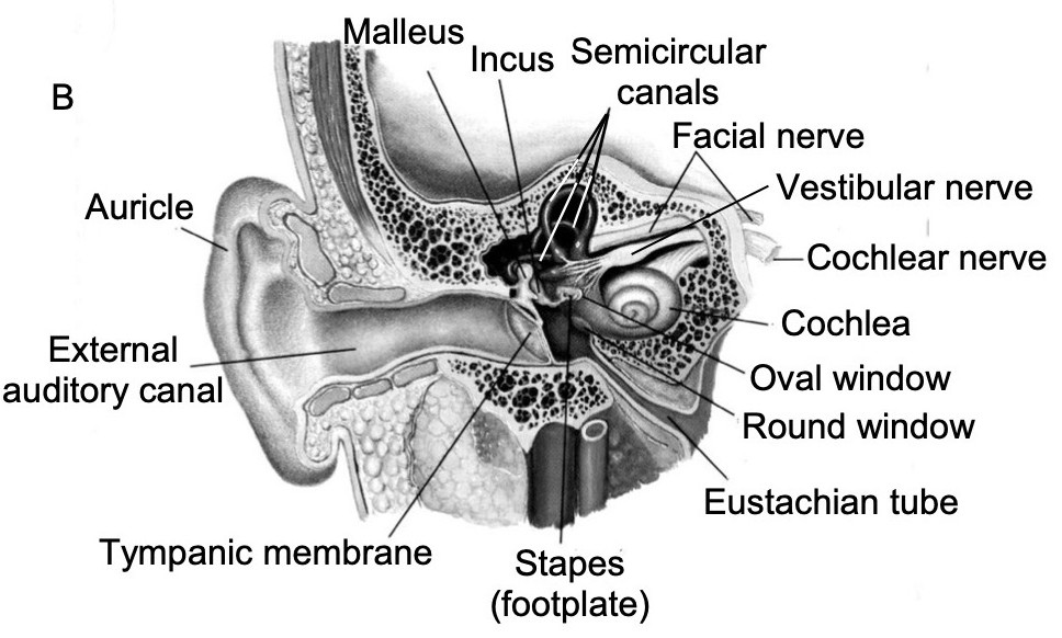
l113_5_cochleaanatomy
 Round window - Wikipedia


/GettyImages-506837613-579d8e123df78c3276da6870.jpg)


Posting Komentar untuk "oval window ear model"